Optical Fiber Integrated High Resolution Unlabeled Differential Microscopic Imaging System
A research team in a Chinese laboratory has proposed the application of traditional fluorescence emission differential (FED) microscopy technology to super-resolution microscopic imaging of non-fluorescent-labeled samples, using the developed fiber mode selection coupler (MSC) to replace the complex spatial light path in the traditional FED microscopy system. A super resolution microscopic imaging system with simple and compact structure, strong ability to resist external interference, and universal application to non-fluorescent labeled samples have been successfully developed.
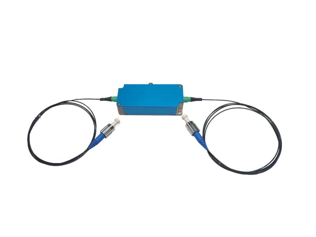
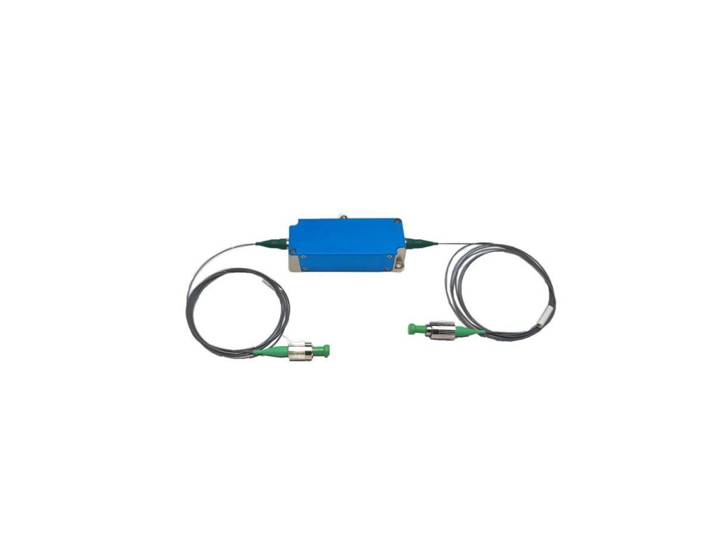
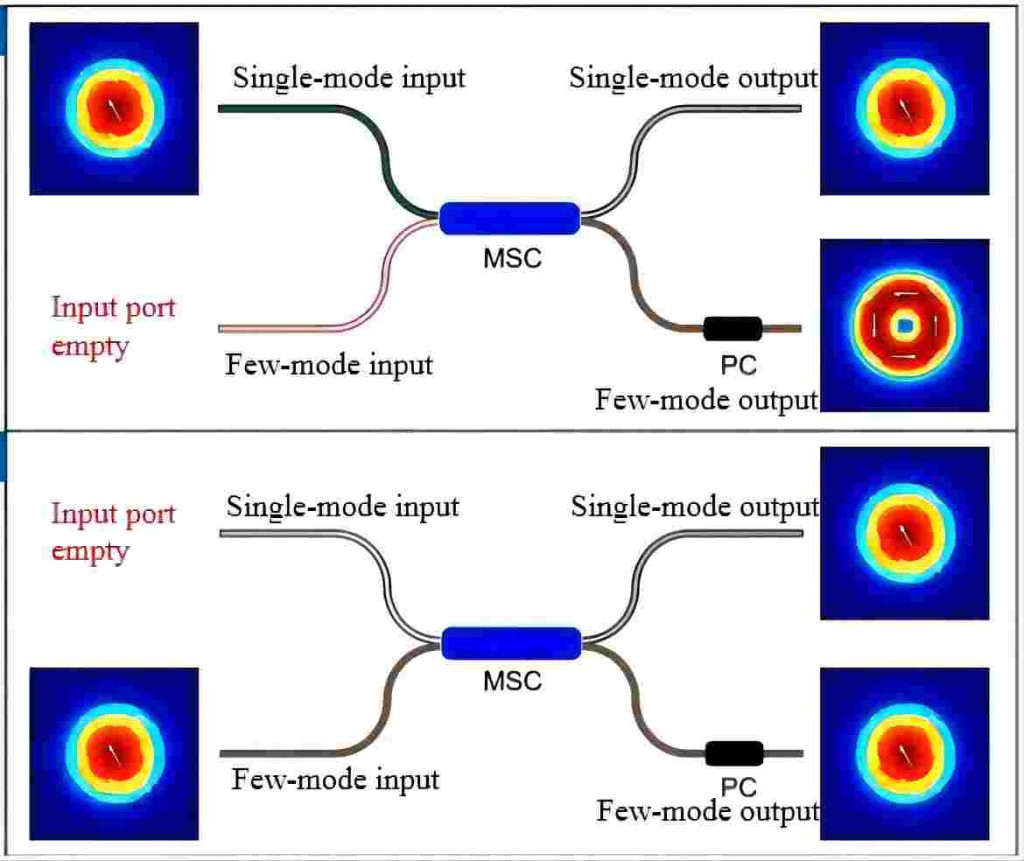
The developed fiber mode selective coupler (MSC) has a feature that whether the excited light is input from the MSC’s single-mode fiber input or from the few-mode fiber input, most of the optical power is coupled to the few-mode fiber output. The difference is that when the excitation light is input from the single-mode fiber, the output end of the few-mode fiber is excited by the circular distribution of LP11 mode; When the excitation light is input from the few-mode fiber, the output of the few-mode fiber is excited by the ordinary Gaussian distribution LP01 mode. The input and output characteristics of optical fiber MSC are shown in Figure 1.
Based on the characteristics of the above fiber MSC, combined with the fiber acousto-optic modulator (AOM) switch which can be switched at high speed, the solid circular optical field and the ring optical field can be time-shared at the few-mode output port of the MSC. The two forms of light field naturally maintain perfect coaxial transmission characteristics after output at the same fiber port. The light field output from the optical fiber MSC is scanned by the scanning galvanometer, and the beam is reduced by the necessary lens group and finally converged by the microscopic objective into the scanning spot used in the imaging process, and the sample is scanned point-by-point. Through the control of fiber AOM, the same sample is scanned twice with a solid spot and a hollow spot, and after the scanning results are weighted and subtracted, the image with super-resolution details can be obtained. The principle of the FED imaging system based on fiber optic MSC is shown in Figure 2.
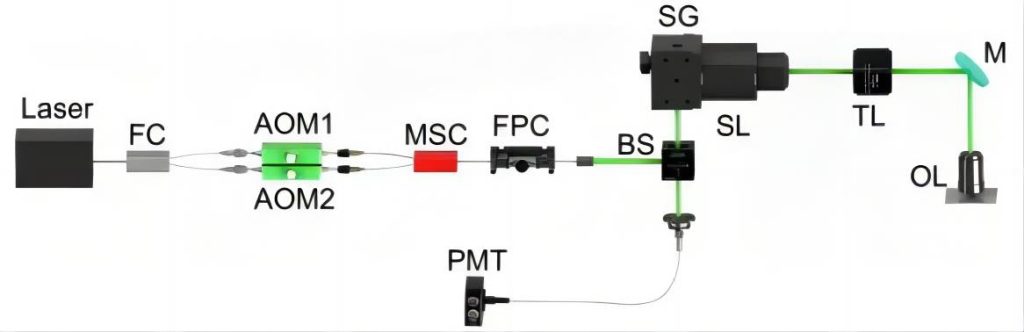
integrated in the fiber optic path; MSC: Fiber mode selective coupler; FPC: Fiber polarization state
controller; BS:Beam spliter; SG:Scanning galvanometer; SL:Scanning lens; TL:Tube lens;
M:Mirror; OL:Objective lens; PMT:Photo-multiplier tube
Figure 2. Schematic diagram of FED imaging system based on fiber optic MSC
Using the imaging system described above, the research team demonstrated the imaging results of a set of gold particle samples with a diameter of about 50 to 150 nanometers, using the impurities to scatter the ring light and the scattering effect on the solid light, the scan results will be different. After weighted subtraction of the scanning results, an image with super-resolution detail information can be obtained. The results demonstrate the improved performance of the system compared to ordinary laser scanning confocal microscopy. If you need more details, please consult the literature. The above is an excerpt from 罗昊,侯梦蝶,徐良,等. 光纤集成化高分辨率无标记差分显微成像系统[J]. 光电工程,2023, 50(12): 230181. doi: 10.12086/oee.2023.230181